Unveiling the Lung Model with Larynx Labeled: Embark on an educational journey into the intricacies of the respiratory system, where a detailed model serves as an invaluable tool for understanding its anatomy, functions, and clinical significance.
Delving into the intricacies of the larynx, we uncover its crucial role in respiration and phonation, meticulously labeled for enhanced comprehension. Prepare to be captivated as we explore the construction, applications, and advancements in lung models, empowering you with a comprehensive understanding of this essential organ system.
Lung Model with Larynx

Lung models with larynx labels are valuable educational tools that provide a comprehensive understanding of the respiratory system. They are typically used in medical schools, science classrooms, and healthcare settings to demonstrate the anatomy and function of the lungs and larynx.
Components and Structures
Lung models with larynx labels typically include the following components and structures:
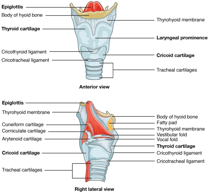
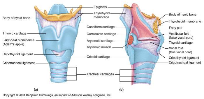
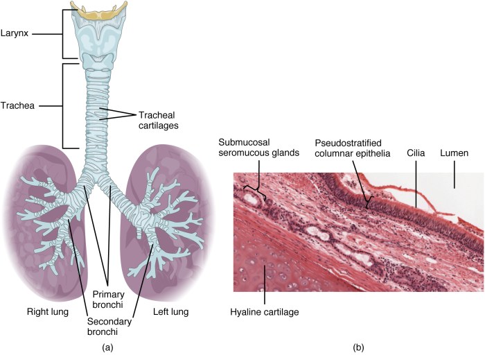
- Larynx: The larynx, also known as the voice box, is located at the top of the trachea. It houses the vocal cords, which vibrate to produce sound.
- Trachea: The trachea is a tube-like structure that carries air from the larynx to the lungs.
- Bronchi: The bronchi are the two main branches of the trachea that enter the lungs.
- Bronchioles: The bronchioles are smaller branches of the bronchi that lead to the alveoli.
- Alveoli: The alveoli are tiny air sacs where gas exchange occurs.
- Diaphragm: The diaphragm is a muscle that separates the chest cavity from the abdominal cavity. It plays a crucial role in breathing.
Anatomy and Structures of the Larynx

The larynx, commonly known as the voice box, is a vital organ located in the upper respiratory tract, just below the pharynx. It plays a crucial role in both phonation (voice production) and respiration.
Key Anatomical Structures of the Larynx
- Epiglottis:A leaf-shaped cartilage that flips down during swallowing, preventing food and liquid from entering the larynx.
- Vocal Cords:Two bands of tissue that vibrate when air passes through them, producing sound.
- Arytenoid Cartilages:Paired cartilages that move the vocal cords, controlling pitch and volume.
Role in Phonation and Respiration
The larynx is essential for speech and singing. The vibration of the vocal cords creates sound waves that resonate in the vocal tract, producing different pitches and tones. Additionally, the larynx acts as a valve, regulating airflow during respiration. The epiglottis closes during swallowing to prevent aspiration of food or liquid into the lungs.
Lung Model Design and Construction

Lung models with larynx labeled are essential tools for medical education and research. They provide a realistic representation of the human respiratory system, allowing students and professionals to visualize and study its complex anatomy. The design and construction of these models require careful consideration to ensure accuracy and detail.
Materials and Techniques
Lung models are typically made from a variety of materials, including plastic, rubber, and latex. These materials are chosen for their durability, flexibility, and ability to accurately replicate the shape and texture of the lungs. The larynx, which is the organ responsible for voice production, is often made from a separate material, such as silicone, to ensure accurate representation of its delicate structures.
Various techniques are employed to construct lung models. One common method is injection molding, where molten plastic is injected into a mold of the desired shape. Another method is handcrafting, where skilled artisans use a variety of tools to shape and assemble the model from individual components.
Importance of Accuracy and Detail
Accuracy and detail are crucial in the design of lung models with larynx labeled. Accurate representation of the anatomical structures allows for precise visualization and study of the respiratory system. This is particularly important for medical students and professionals who need to understand the complex interplay of different structures within the lungs and larynx.
Detailed models provide a more comprehensive understanding of the respiratory system. They can include features such as the bronchial tree, alveoli, and blood vessels, allowing for a thorough examination of the anatomical relationships and functional aspects of the lungs.
A lung model with larynx labeled is a great teaching tool for anatomy students. The larynx is a crucial part of the respiratory system, and understanding its structure and function is essential for students in various fields, including cosmetology. The nd state board of cosmetology requires cosmetology students to have a basic understanding of anatomy, and a lung model with larynx labeled can be a valuable resource for them.
Types of Lung Models
There are various types of lung models available for educational and research purposes. These models differ in their size, complexity, and level of detail.
- Basic models: These models provide a simplified representation of the lungs and larynx, focusing on the main anatomical structures.
- Advanced models: These models offer a more detailed representation, including intricate structures such as the bronchial tree and alveoli.
- Interactive models: These models allow users to manipulate and interact with the model, simulating breathing and other respiratory functions.
- Patient-specific models: These models are created using medical imaging data from individual patients, providing a personalized representation of their respiratory system.
The choice of lung model depends on the specific educational or research needs. Basic models are suitable for introductory courses, while advanced and interactive models are more appropriate for advanced studies and research.
Applications and Uses of Lung Models
Lung models with larynx labeled are valuable educational and research tools.
Educational Applications, Lung model with larynx labeled
- Medical Training: These models aid medical students and healthcare professionals in understanding the complex anatomy and physiology of the respiratory system. They provide a tangible and interactive way to study the larynx, trachea, and lungs.
- Patient Education: Lung models can help patients visualize their respiratory system and comprehend the impact of various respiratory conditions. This enhanced understanding promotes informed decision-making and improves patient adherence to treatment plans.
Research Applications
- Respiratory Disorders: Researchers use lung models to investigate the mechanisms and progression of respiratory diseases. By simulating various conditions, they can evaluate the effectiveness of treatments and develop new therapeutic strategies.
- Surgical Planning: These models assist surgeons in preoperative planning by providing a detailed representation of the patient’s anatomy. This visualization helps optimize surgical approaches, minimize risks, and improve patient outcomes.
Enhancing Understanding
Lung models facilitate a deeper comprehension of the respiratory system. They allow students and researchers to visualize the intricate structure of the larynx and lungs, examine the flow of air during respiration, and explore the interactions between different respiratory components.
Advanced Features and Technologies: Lung Model With Larynx Labeled

Lung models with larynx labeled are continuously evolving, incorporating advanced technologies to enhance their accuracy, functionality, and educational value. These technologies offer unique advantages in the design and construction of lung models, enabling researchers, educators, and medical professionals to explore the complexities of the respiratory system in unprecedented ways.
One significant advancement is the use of 3D printing technology. 3D printing allows for the creation of highly detailed and anatomically accurate lung models, complete with intricate structures such as the larynx, bronchi, and alveoli. These models can be customized to represent specific patient anatomies or disease conditions, providing personalized training and educational experiences.
Benefits of Advanced Technologies
- Enhanced Accuracy:Advanced technologies enable the creation of lung models with greater anatomical accuracy, capturing the intricate details of the respiratory system.
- Customization:3D printing and other technologies allow for the customization of lung models, tailoring them to specific research or educational needs.
- Interactive Simulations:Virtual reality and augmented reality technologies can create immersive simulations, allowing users to interact with lung models and explore their functionality in a dynamic environment.
Limitations of Advanced Technologies
- Cost:Advanced technologies can be expensive, limiting their accessibility for some institutions and individuals.
- Technical Expertise:Using and maintaining advanced lung models may require specialized technical expertise, which can be a barrier to their widespread adoption.
- Accuracy Validation:While advanced technologies enhance accuracy, it is crucial to validate the models against real-world data to ensure their reliability.
Examples of Innovative Lung Models
Several innovative lung models incorporate advanced features and technologies. For instance, the “Virtual Lung” project developed by the University of California, San Francisco, uses virtual reality to create a realistic simulation of the respiratory system. Users can navigate through the lung model, interact with its structures, and simulate various respiratory conditions.
Another example is the “3D Printed Lung Model” developed by the University of Pittsburgh. This model is 3D printed using patient-specific data, providing a personalized and highly accurate representation of the individual’s respiratory anatomy.
Comparison with Other Respiratory Models

Lung models with larynx labeled provide valuable insights into the anatomy and function of the respiratory system, but they are not the only tools available for studying this complex system. Other types of respiratory models, such as chest X-rays and CT scans, offer unique perspectives and can be used in conjunction with lung models to provide a comprehensive understanding of the respiratory system.
Chest X-rays are a common and widely available imaging technique that provides a two-dimensional view of the chest cavity. They can be used to detect abnormalities in the lungs, such as pneumonia, lung cancer, and heart failure. However, chest X-rays do not provide as much detail as CT scans and can be difficult to interpret in some cases.
CT scans are a more advanced imaging technique that provides detailed cross-sectional images of the chest cavity. They can be used to diagnose a wide range of respiratory conditions, including lung cancer, emphysema, and pulmonary embolism. CT scans provide more information than chest X-rays, but they are also more expensive and time-consuming to perform.
Lung models with larynx labeled can be used to complement other respiratory models by providing a three-dimensional representation of the respiratory system. This can be helpful for understanding the spatial relationships between different structures and for visualizing how the respiratory system functions as a whole.
Lung models can also be used to demonstrate respiratory procedures, such as intubation and tracheotomy.
The choice of which type of respiratory model to use depends on the specific application. Chest X-rays are a good option for screening for respiratory conditions, while CT scans are more useful for diagnosing and monitoring respiratory diseases. Lung models with larynx labeled can be used to supplement other respiratory models by providing a three-dimensional representation of the respiratory system.
Detailed FAQs
What is the purpose of a lung model with larynx labeled?
Lung models with larynx labeled serve as educational and research tools, providing a detailed representation of the respiratory system’s anatomy, particularly focusing on the larynx and its structures.
How are lung models with larynx labeled constructed?
These models are typically made using various materials such as plastic or resin, ensuring accuracy and durability. Advanced techniques like 3D printing and virtual reality are also employed to enhance their realism and functionality.
What are the benefits of using lung models with larynx labeled?
These models offer a tangible and interactive way to study the respiratory system, facilitating a deeper understanding of its structures, functions, and potential disorders. They are particularly valuable in medical education, patient education, and research.